
Brain Tumor Surgery in Patna
Nirog Hospital offers state-of-the-art neurosurgical interventions using Neuronavigation, high-end operating microscopes, and advanced neuromonitoring to ensure maximum safety and tumor resection.
Top Neurosurgeons
Experienced specialists on board
Modern Technology
Neuronavigation & Micro-surgical OTs
Cashless & TPA
CGHS, Ayushman & Major TPAs
24x7 Emergency
Trauma, ICU & Neurosurgical care
Understanding Brain Tumors: Overview, Development & Growth
A brain tumor is an abnormal growth of cells in or around the brain that can increase pressure inside the skull and affect normal brain function. Brain tumors may be primary (originating in the brain) or secondary (spreading from cancer in another part of the body).
Brain tumors can be benign (non-cancerous) or malignant (cancerous). As they grow, they may put pressure on nearby brain tissues and affect normal brain function. Early diagnosis, accurate evaluation, and timely treatment are important for achieving the best possible outcomes.
At Nirog Hospital, Patna, we believe early diagnosis is essential for effective brain tumor treatment. Our experienced specialists use advanced diagnostic technology and detailed neurological evaluations to accurately diagnose the condition and develop a personalized treatment plan for every patient.
Brain Anatomy & Tumor Development
The human brain is divided into distinct regions. Learn how tumors in different locations affect critical neurological and bodily functions.
Cerebrum (Main Brain)
Largest Region
Controls voluntary movements, speech, sensory processing, emotions, memory, and cognitive skills.
Tumors here lead to muscle weakness/paralysis on one side of the body, sensory losses, memory slip-ups, or sudden localized seizures.
Cerebellum (Lower Back)
Coordination Center
Coordinates voluntary muscle activity, fine motor control, maintaining body posture, and physical balance.
Tumors in this zone impair stability directly, causing staggering gait (unsteady walking), dizziness, and loss of hand coordination.
Brain Stem (Lower Base)
Life Regulation
Connects the brain to the spinal cord, regulating critical automated life functions like breathing, heart rate, and blood pressure.
Even tiny growths in this confined base region can be life-threatening, causing double vision, swallowing difficulties, or breathing issues.
Different Types of Brain Tumors We Treat
Brain tumors are classified based on their cellular origin and how aggressively they grow.
Benign vs. Malignant
ClassificationBenign tumors grow slowly, have clear borders, and rarely invade adjacent tissues. However, they can still cause severe pressure symptoms inside the skull. Malignant tumors contain cancerous cells, lack clear borders, grow aggressively, and infiltrate nearby brain matter, needing prompt surgical removal and chemotherapy/radiation.
Gliomas & Astrocytomas
Primary TumorsOriginating in the supportive glial cells, gliomas represent a major portion of primary brain malignancies. These include Glioblastoma Multiforme (GBM) – a highly aggressive Grade IV tumor. Safe resection assisted by advanced neuromapping is vital to optimize patient survival and lifestyle.
Meningiomas & Pituitary
Membrane & GlandularMeningiomas arise from the brain's protective membranes and are often benign, though they can grow large. Pituitary tumors grow in the hormone-regulating gland at the base of the skull, causing hormonal imbalances and visual changes as they press on the optic nerve.
Recognizing Early & Advanced Symptoms
Symptoms can vary from general signs of pressure inside the skull to specific deficits caused by tumor localization.
Early Warning Signs
- Persistent headaches, especially worse in the morning or during coughing.
- Unexplained, sudden projectile vomiting and frequent nausea.
- Gradual changes in vision, such as double vision or blurred sight.
- Mild coordination problems, dizziness, or loss of balance.
Advanced Symptoms
- Sudden seizures (fits) in patients with no history of epilepsy.
- Localized weakness or numbness in arms, legs, or one side of the face.
- Difficulty speaking, finding words, or understanding spoken language.
- Significant personality changes, confusion, memory loss, or severe mood swings.
What Causes Brain Tumors & Who is at Risk?
Brain tumors develop from DNA anomalies. Explore the key risk factors and clinical causes below.
DNA Mutations & Genetic Factors
Like most cell-growth disorders, primary brain tumors develop when errors (mutations) occur in the DNA of healthy cells. These genetic errors instruct the cells to divide rapidly and survive when normal cells would die, forming a growing mass.
Inherited genetic syndromes such as Neurofibromatosis Type 1 and 2, Li-Fraumeni syndrome, and Turcot syndrome are known to significantly increase the risk of developing brain tumors.
Brain Tumor Stages & WHO Grades
Unlike other cancers, brain tumors are categorized by grades (I to IV) based on cell appearance under a microscope and growth rate, rather than bodily spread.
| Grade | Growth Rate | Characteristics | Typical Types |
|---|---|---|---|
| Grade I | Slow-growing | Non-cancerous cells, clear borders; surgery is often curative. | Pilocytic Astrocytoma |
| Grade II | Relatively slow | Slightly abnormal cells; can spread to nearby tissues and recur. | Diffuse Astrocytoma |
| Grade III | Rapid-growing | Malignant cells that actively reproduce and infiltrate healthy brain tissue. | Anaplastic Astrocytoma |
| Grade IV | Highly aggressive | Aggressive cancer; rapidly invades tissue, forms new blood vessels. | Glioblastoma (GBM) |
When Should You Consult a Neurosurgeon Immediately?
If you or your loved ones experience a sudden onset of seizures, unexplained progressive numbness in the limbs, persistent morning headaches accompanied by vomiting, or rapid changes in visual field and speech, consult a neurosurgeon immediately for evaluation.
Advanced Diagnosis & Brain Tumor Testing
We combine advanced neuroimaging technology with specialist consultations to accurately diagnose brain tumors.
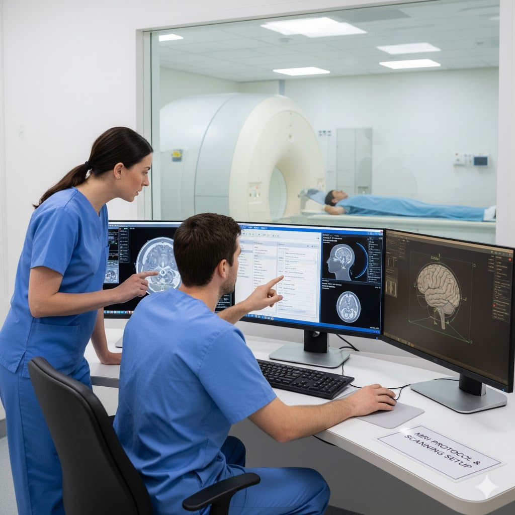
High-Resolution MRI Brain Scan
Uses strong magnetic fields and radio waves to generate slice-by-slice clinical images of the brain structure. Contrast dye is used to highlight the tumor borders clearly, assisting the neurosurgeon in precise mapping before any surgery.
Comprehensive Treatment Options
Treatment depends on the type, size, location of the tumor, and the patient's overall health. Click on any card below to expand advanced clinical details.
Medicines
Corticosteroids to reduce swelling and edema around the tumor. Anti-seizure medications to prevent fits and control neurological stability.
Dexamethasone is commonly used to manage increased pressure. Anticonvulsants (e.g., Levetiracetam) are crucial to secure electrical pathways in the brain.
Surgery
Neurosurgical removal (Craniotomy) to safely resect as much of the tumor as possible. This is the primary treatment for most primary brain tumors.
Utilizes computer-assisted neuronavigation, intraoperative MRI, and cortical mapping to achieve maximal safe tumor resection while protecting vital speech/motor regions.
Radiation
High-energy beams (X-rays or protons) to target and destroy remaining tumor cells after surgery, or as primary treatment for deep-seated tumors.
Includes Intensity-Modulated Radiation Therapy (IMRT) and stereotactic radiosurgery (SRS/Gamma Knife) to target cancer cells with sub-millimeter precision.
Chemotherapy
Uses powerful drugs (oral or intravenous, such as Temozolomide) to stop the growth of malignant cells, often combined with radiation therapy.
Uses drugs capable of crossing the blood-brain barrier. It acts globally to stop cell replication, often scheduled in cycles following surgical resection.
Brain Tumor Surgery: How it works?
Traditional Craniotomy
Surgically opening the skull cavity to access and remove as much of the tumor mass as possible.
Awake Surgery
Performed conscious with mapping to protect and monitor crucial speech and motor pathways.
Endoscopic Surgery
Minimally invasive access through nasal cavity pathways, typically for pituitary tumors.
Stereotactic Biopsy
A minimally invasive needle procedure guided by 3D computer systems for precise sampling.
Precision Technology for Safe Brain Surgery
We use advanced surgical technologies to maximize tumor resection while protecting healthy brain tissue.
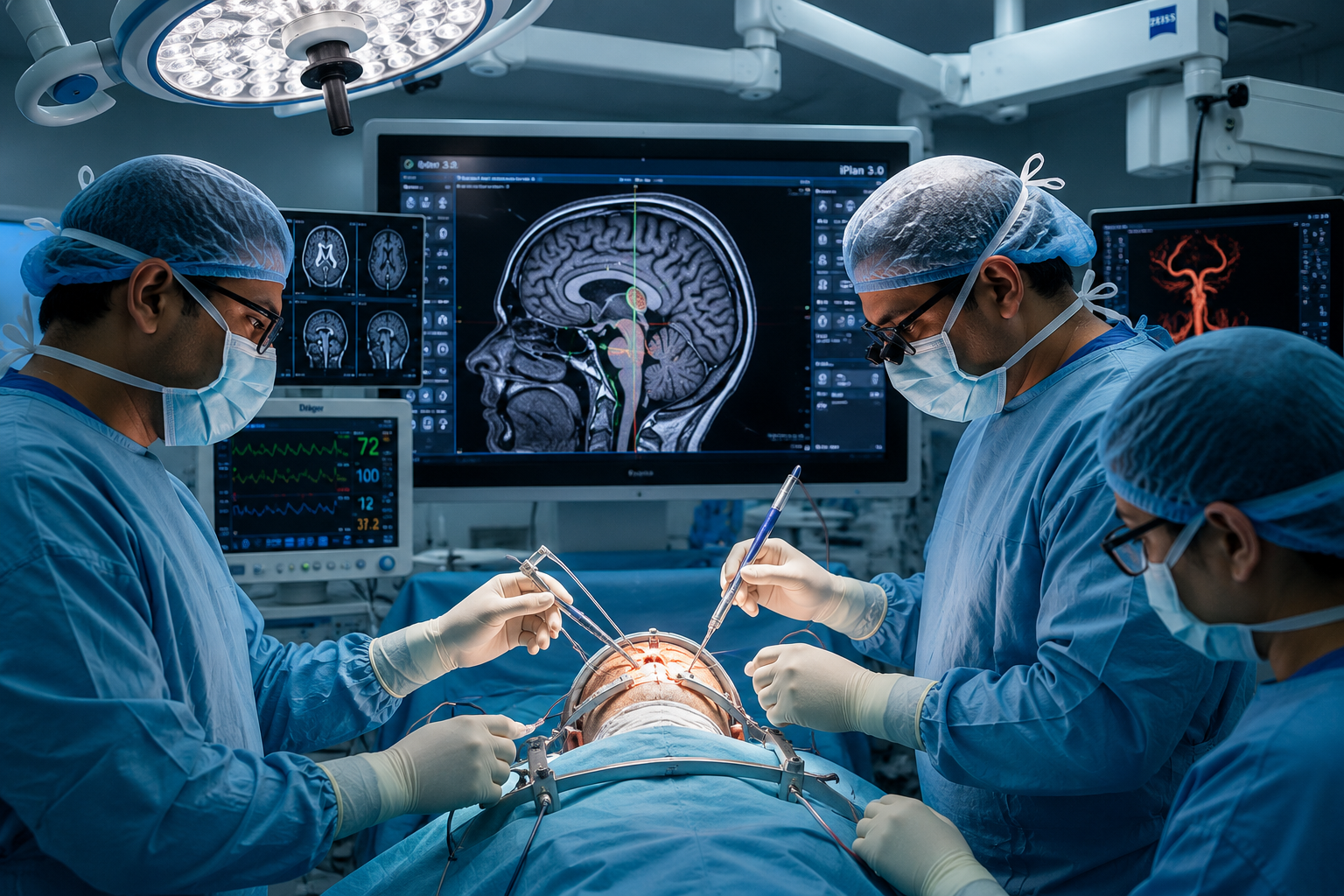
3D Neuronavigation System
Sub-millimeter GPSActs like a GPS system for the brain, allowing neurosurgeons to track and target tumors with sub-millimeter precision.
Surgical Microscope
Stereoscopic ViewProvides stereoscopic magnification and illumination, helping surgeons distinguish healthy brain tissue from tumors.
Neuromonitoring (IONM)
Nerve ProtectionMonitors speech and motor pathways in real-time, helping prevent damage to vital brain functions during resection.
Preparing Checklist
Essential preoperative readiness steps for patient safety and successful surgery:
Surgical Procedure Path
Anaesthesia & Position
General anaesthesia is administered, and the patient is positioned securely to expose the surgical site with neuro-navigation sensors calibrated.
Accessing the Tumor
A precise craniotomy is performed using 3D coordinates. A temporary bone flap is removed, exposing the protective meninges layer of the brain.
Resection & Closing
The tumor is resected under microscope magnification. The bone flap is secured back with micro-plates, and the scalp tissues are closed carefully.
Postoperative Recovery & Rehabilitation
Recovery after brain surgery is a gradual process that involves monitoring, physical therapy, and follow-up care.
Hospital Stay & Home Recovery
Patients spend the first 24 to 48 hours in the ICU for close neurological monitoring. The typical hospital stay is 4 to 7 days, followed by home rest. Regular wound care and follow-up MRI scans are key to monitoring healing.
Neurological Rehabilitation
Our rehabilitation team includes speech therapists to address communication issues, physical therapists to rebuild motor strength, and occupational therapists to help patients regain independence in daily activities.
Risks, Complications & Long-term Prognosis
Brain surgery is a complex procedure. While potential risks like brain swelling, infections, or seizures exist, we utilize advanced neuromonitoring and surgical mapping to minimize these concerns.
Prognosis depends heavily on the tumor's grade, type, location, and the patient's general health. Slow-growing benign tumors often achieve complete recovery, whereas malignant variants require a multi-disciplinary management path.
Swelling Control
Brain swelling (edema) is strictly managed using corticosteroids and hyperosmolar therapy.
Infection Shield
Surgical site infections are prevented using strict sterile modular OTs and targeted prophylactic antibiotics.
Seizure Prevention
Postoperative seizures are monitored and mitigated with customized anticonvulsant protocols.
Brain Tumor Surgery Cost & Cashless Facilities
Nirog Hospital provides affordable neurosurgery options with transparent pricing and insurance support.
Cost Estimation
Neurosurgery costs depend on factors such as tumor complexity, the type of surgery, hospital stay duration, and diagnostic requirements. We offer competitive rates and transparent billing to help manage expenses.
Cashless & TPA Partners
We work with major insurance providers and third-party administrators (TPA) to offer cashless options. Nirog Hospital also accepts government schemes such as Ayushman Bharat (AB-PMJAY) and CGHS to support patients.
Why Choose Nirog Hospital for Brain Tumor Surgery in Patna?
Experienced Neurosurgeons
Top-tier specialists with years of complex brain tumor resection experience.
Dedicated Neuro-ICU
Round-the-clock intensive care and critical support by trained staff.
Advanced Modular OT
Modular operating suites with strict infection control measures.
Neuronavigation & IONM
Real-time GPS mapping and nerve monitoring during resection.
Ayushman & TPA Accepted
Cashless treatment under major government schemes and corporate insurances.
Transparent Costing
High-quality care with clear, affordable package structures.
Our Specialized Neuro Care Facilities
Designed to support postoperative care and surgical safety.
Neuro-Intensive Care (ICU)
Monitored beds with specialist nursing care.
Advanced Operation Theatres
Neurosurgical suites equipped with microscopes.
24x7 Diagnostics & Pharmacy
In-house laboratories, MRI, and pharmacy support.
Patient Video Stories
Watch real stories from our patients about their healing journey and expert insights from our doctors.
Patient Testimonials & Recovery Stories
Real recovery journeys of brain tumor surgery patients treated at Nirog Hospital Patna.
"My brother was diagnosed with a brain tumor after sudden seizures. Dr. and team performed microscopic craniotomy at Nirog Hospital. He made a complete neurological recovery."
Amit Roy
Patna, Bihar"100% cashless brain tumor surgery under Ayushman Bharat scheme. The neuronavigation system and 24x7 Neuro ICU care gave us immense confidence."
Vikram Kumar
Bhagalpur, Bihar"We came from outstation for skull base tumor treatment. The hospital supported us with priority admission, lodging guidance, and post-surgery rehabilitation."
Sangeeta Devi
Siwan, BiharFrequently Asked Questions
Ready to get started?
Schedule a consultation with our experienced neurosurgery specialists today for a personalized assessment.